
What is the Human Nervous System?
https://bodytomy.com/human-nervous-system-diagram
Reading this article will be like reading about the human nervous system from your science textbook as this article carries a labeled human nervous system diagram to help you understand its design and working. For me, diagrams and illustrations that textbooks carried, were always of great help in understanding the topic. It was the labeled animal cell diagram that ‘magnified my image’ of the microscopic cell. It was the skeletal system diagram that helped me understand the “skeleton” of human bodies. And it was when I first studied the diagram of a human heart, I realized that the heart is not exactly heart-shaped!
Coming back to the point, let me start explaining the human nervous system function and parts with the help of a labeled diagram. Before going to the details of the structure and functioning of the human nervous system, you should know what the nervous system actually is and what it does. The nervous system is a network of special cells, neurons and ganglia, that work together to carry out the transmission and reception of signals between different parts of our body. The signals are transmitted in the form of electrochemical waves or chemicals. Before you proceed to understand the human nervous system function and parts, you might like to go through some human nervous system facts. So here are some interesting facts about the nervous system. Let’s come to answer how the nervous system works.
Neurons
Neuron can be considered as the basic unit of the nervous system, which processes and transmits information by means of electrochemical signals. Sensory neurons respond to external stimuli that affect the sensory organ cells. Motor neurons, on receiving signals from the central nervous system, bring about responses at the target organs. Interneurons act as the connectors between neurons. Neurons are of different shapes and sizes and their complex interconnections add to the complexity of the nervous system. The human brain contains 86.1 billion neurons.
Glia
Glia or glial cells, as they are called, are non-neural cells that play a vital role in maintaining homeostasis and protecting the brain’s neurons. The glial cells surround the neurons to hold them in place, supply them with oxygen and nutrients, isolate the neurons from one another and remove dead neurons. The human brain contains about 84.6 billion glia; that’s almost equal to the number of neurons it contains.
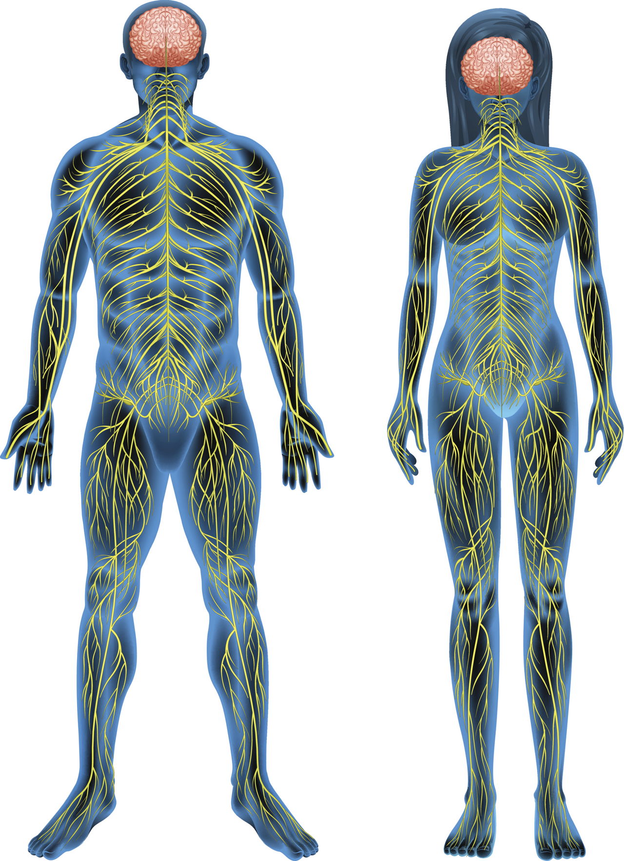
The human nervous system can be divided into two parts, central and peripheral. The central nervous system (CNS) consists of the brain and the spinal cord, while the peripheral nervous system (PNS) consists of sensory neurons, ganglia (clusters of neurons) and nerves. Here is a diagram that you can refer to, while you read about the human nervous system function and parts.
Central Nervous System

The central nervous system coordinates the functioning of all parts of the human body and is the largest part of the nervous system. It is enveloped by a set of membranes known as meninges that protect the brain and the spinal cord. They also have their own protective covers! The skull protects the brain while the vertebrae and spinal cavity shield the delicate spinal cord. To be precise, the brain is placed in the cranial cavity and the spinal cord in the spinal cavity. Let’s take a closer look at the central nervous system parts and functions.
Brain
The brain is the center of the human nervous system and is a highly complex organ. Even a brainiac won’t find it very easy to understand the brain! The human brain is about three times larger than the brain of a typical mammal.
The brain can be said to have three main parts, the brain stem, the cerebrum and the cerebellum. The cerebrum is associated with information storage and processing; the cerebellum is responsible for balance, posture and coordination of movements; and the brain stem plays a vital role in controlling breathing and heart rate along with some other important body processes. Along with the skull, the brain is also protected by the cerebrospinal fluid in which it is suspended. It’s strange yet true that the brain floats in a fluid!
For a detailed study of brain anatomy and its functions, read: diagram of the brain and its functions.
Spinal Cord
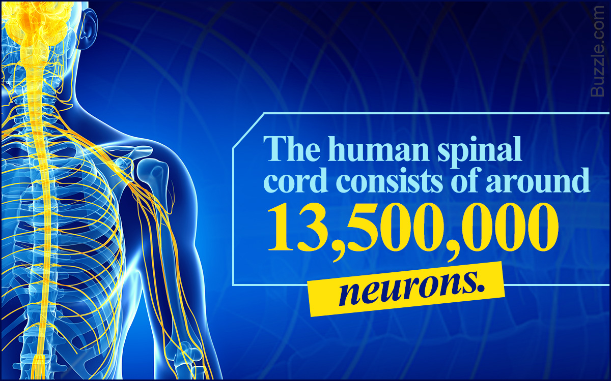
The spinal cord is a long tubular structure composed of nervous tissue and support cells. It is around 45 cm long in men and 43 cm long in women. It extends from the brain up to the space between the first and the second lumbar vertebrae. It transmits neural signals between the brain and other body parts. It is the spinal cord which connects the brain and the peripheral nervous system. It consists of about 13,500,000 neurons.
Peripheral Nervous System
How does the central nervous system communicate with the other body organs? It is through the peripheral nervous system.
Functionally, the peripheral nervous system can be divided into two
parts; the somatic nervous system and the autonomic nervous system, the
somatic nervous system is responsible for bodily activities that are
under conscious control. For example, controlling body movements and
receiving external stimuli. The autonomic nervous system is further
divided into sympathetic, parasympathetic and enteric nervous systems.
The sympathetic nervous system governs the bodily responses to impending
dangers, while the parasympathetic system is responsible for bodily
actions that help in relaxation of body organs that are hyperactive. The
enteric nervous system specifically manages the functioning of the
digestive system. Let’s take a close look at what constitutes the
peripheral nervous system.
Nerves
Cable-like in appearance, the nerves serve as paths for the transmission of nerve impulses along axons. Nerves are found only in the peripheral nervous system. Depending on the direction of the signals they conduct, they are classified into afferent and efferent nerves. The afferent ones conduct signals from sensory neurons to the central nervous system, while the efferent ones conduct signals from the central nervous system along motor neurons to muscles or glands. There are some nerves which can function like both afferent and efferent ones. They are called mixed nerves. Let’s look at the major nerves in the human nervous system.
Musculocutaneous Nerve
It is a part of the
brachial plexus. It runs through the neck, the armpit area and ends in
the arm. It serves the bicep muscles and the skin of the forearm.
Radial Nerve
It is also a part of the brachial
plexus. It supplies the triceps brachii muscle of the arm and a part of
the forearm along with its associated joints and skin.
Median Nerve
It is one of the main nerves that
originate from the brachial plexus. It runs down the arm and enters the
forearm. The median nerve is the only nerve passing through the carpal
tunnel.
Iliohypogastric Nerve
It originates from the
first lumbar nerve and supplies the abdominal muscles along with skin of
some parts of the abdominal wall.
Obturator Nerve
It is a mixed nerve that arises
from the lumbar plexus. It supplies the adductor, gracilis and obturator
externus muscles. It also supplies a part of the skin of the thigh, hip
and knee joints.
Genitofemoral Nerve
It arises in the lumbar
plexus and bifurcates into two branches, namely, genital and femoral.
Its branches run through the skin of the scrotum and to the upper
anterior aspect of the thigh.
Ulnar Nerve
It runs near the ulna bone and is
directly connected to the little finger and half of the ring finger. It
supplies the tips of these fingers and the far back of the fingernail
beds. It is the largest nerve which is unprotected by muscle or bone.
Common Peroneal Nerve
Also known as the common
fibular nerve, it is half the size of the tibial nerve and originates
from the branches of the lumbar and sacral nerves. It runs obliquely
along the side of the depression at the back of the knee joint to the
head of the calf bone.
Deep Peroneal Nerve
Also known as the deep
fibular nerve, it originates at the bifurcation of the common peroneal
nerve, comes above the middle of the leg and then to the front of the
ankle joint. The deep peroneal nerve supplies muscular branches to some
parts of the leg and the ankle joint.
Superficial Peroneal Nerve
It supplies the
peroneus longus, a muscle in the lateral compartment of the leg and the
peroneus brevis, a smaller muscle lying under the peroneus longus. This
nerve supplies musclular branches to the longus and the brevis muscles
and cutaneous filaments to the skin of the lower part of the leg.
Tibial Nerve
The tibial nerve is a branch of the
sciatic nerve. It passes through the depression at the back of the knee
joint, where it gives off an articular branch to the knee joint and a
cutaneous branch that becomes the sural nerve.
Saphenous Nerve
It is the largest cutaneous
branch of the femoral nerve. It supplies cutaneous branches to the skin
of the leg and foot in the region between the knee and the ankle.
Sciatic Nerve
Also known as the ischiatic nerve,
the sciatic nerve is a nerve fiber that begins in the lower back and
ends in the lower limb. It supplies the skin of the leg and the muscles
of the leg, foot and back of the thigh.
Pudental Nerve
Originating in the sacral region
of the spinal cord, it is formed from the second, third and fourth
sacral nerves. It is located in the pelvic region and it supplies the
external genitalia of both men and women.
Femoral Nerve
It is located inside the leg and
supplies muscles that help in bending and straightening the leg. It is
the largest branch of the lumbar plexus.
Subcostal Nerve
It is the vertical branch of the
12th thoracic nerve and supplies some parts of the abdominal muscles. It
supplies branches to the skin of the lower abdominal wall and the
gluteal region. It passes along the border of the 12th rib.
Intercostal Nerves
The ventral branches of the
thoracic nerves are known as intercostal nerves. The first two
intercostal nerves supply fibers to the upper limb, the next four, to
the thorax and the lower five to the thorax and abdomen.
Plexus
The literal meaning of plexus is network. In human beings, rather all vertebrates, the area where nerves branch and rejoin, is known as a plexus. The four main nerve plexuses in the human body are cervical plexus, brachial plexus, lumbar plexus and sacral plexus. Here is a brief description of each of them.
Cervical Plexus
The anterior branches of the
upper four cervical nerves form a plexus in the neck region, and it is
known as the cervical plexus. The nerves of the cervical plexus supply
the back of the head, the neck and the shoulders.
Brachial Plexus
The arrangement of nerve fibers
formed by the ventral rami of the lower cervical and upper thoracic
nerve root, precisely between the nerve roots of the 5th cervical and
1st thoracic vertebra, is known as the brachial plexus. It runs through
the neck, the armpit region and then into the arm.
Sacral Plexus
This plexus supplies nerves to the
posterior thigh, lower leg, foot and a part of the pelvis. It is formed
by the 4th and 5th lumbar nerves and the 1st, 2nd and 3rd sacral nerves.
Nerves of the sacral plexus join to form a flattened band, which later
forms the sciatic nerve.
Lumbar Plexus
Formed by the ventral divisions of
the first four lumbar nerves, the lumbar plexus is located in the lumbar
region of the body. It passes through psoas major, where it supplies
some smaller motor branches. Some of its larger branches run through the
pelvic area.
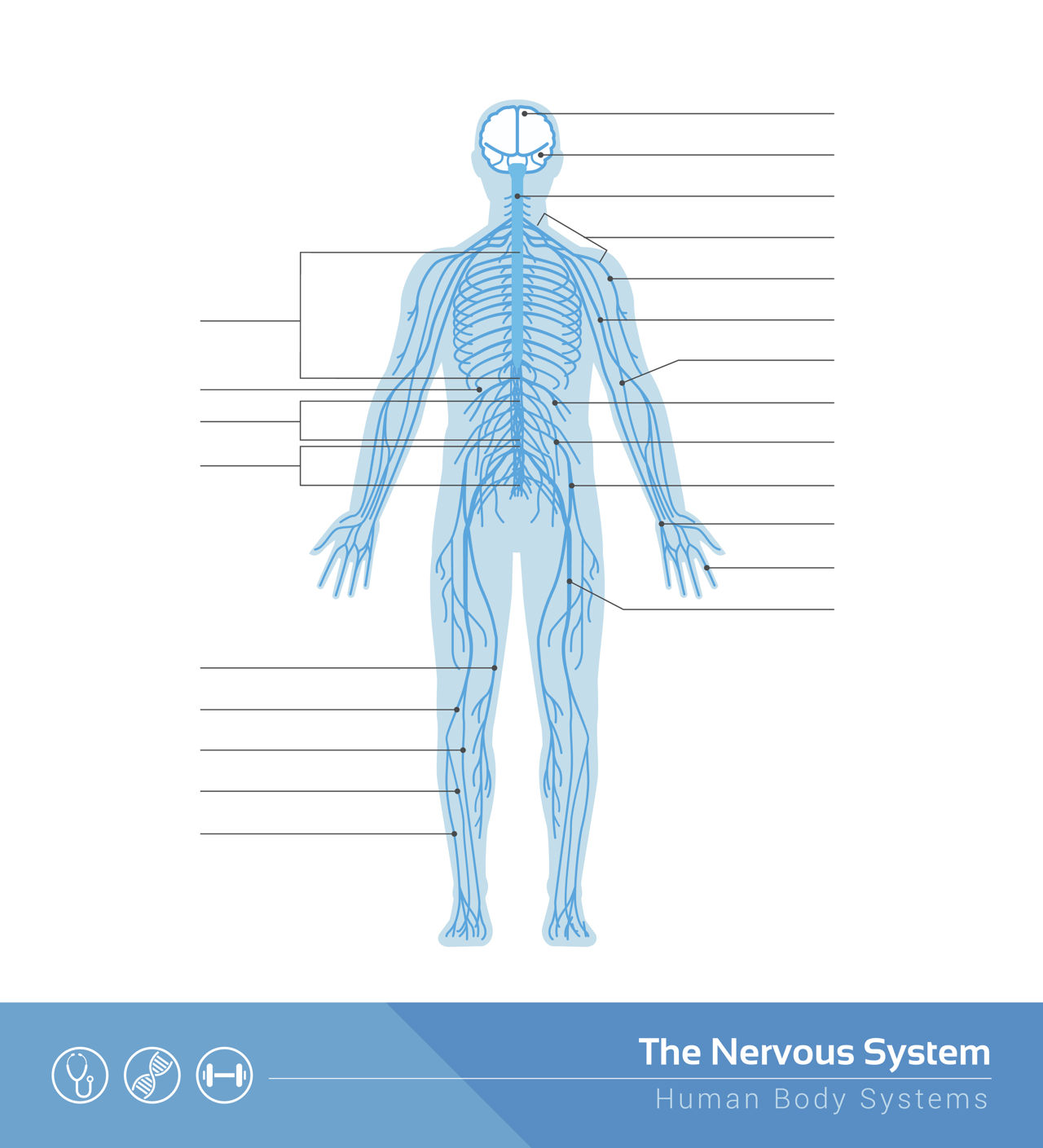
This was an overview of the human nervous system function and structure along with a labeled diagram. I hope it helped you understand the nervous system function and parts and also gave you that ‘textbook feel’ while reading. Here is something that will add to that feeling; a human nervous system diagram to label. Download the image (right click and save) and try labeling all the nervous system parts that you just studied. Before you begin, you can take a quick peek at the already labeled diagram given above and see if you get the labels right!
I will conclude the article with what my teacher had said after she finished her lessons on human nervous system. She had said, “The nervous system, rather any human body system is definitely not very easy to understand. Studying human anatomy is in fact, understanding the most complex and intelligently crafted machines. And understanding complexities can never be simple. But making the complex appear simple is what only God can do.”


Comments
Post a Comment